In‑Vitro Data That Moves Fast, Like Your Skin
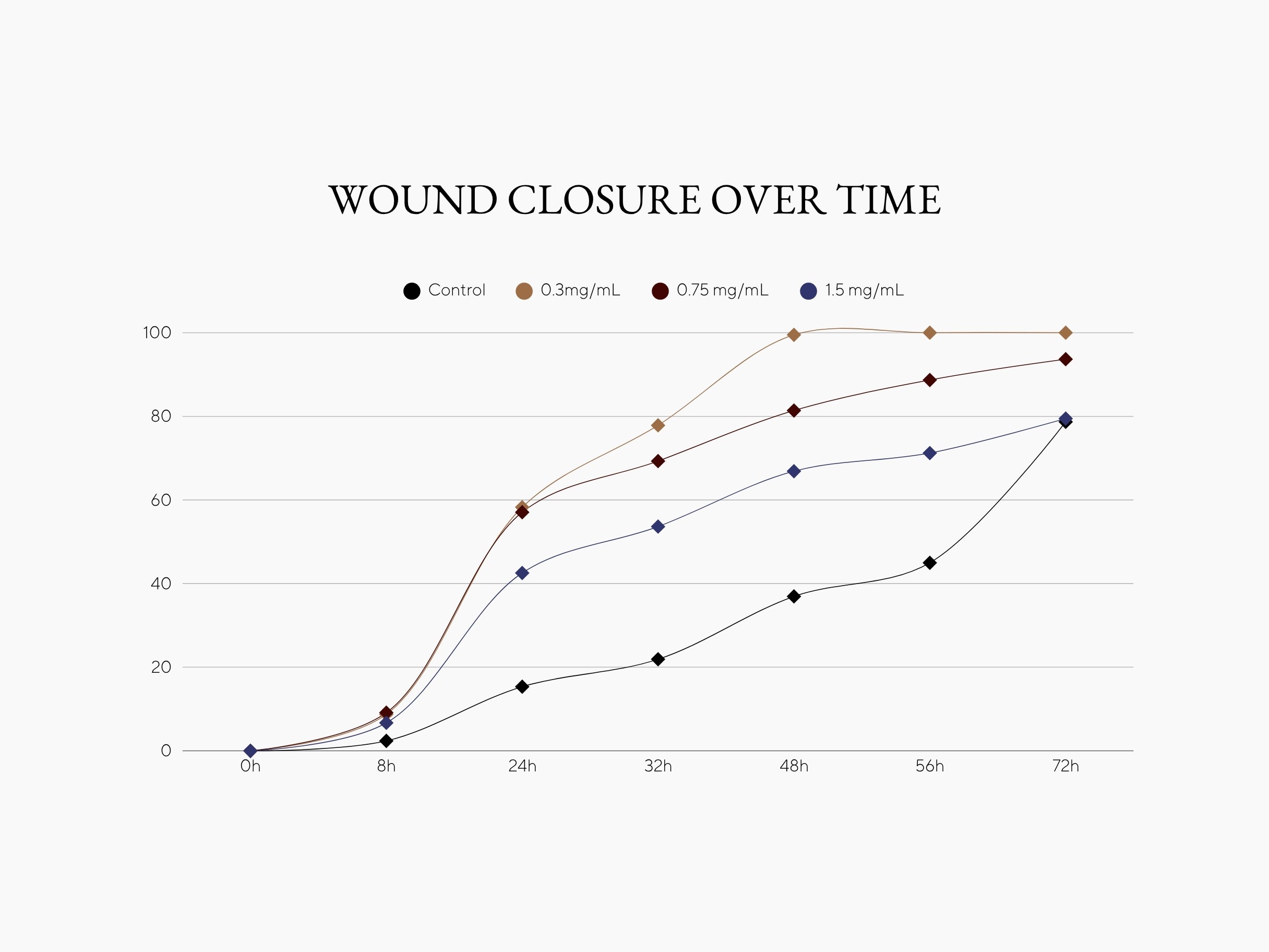
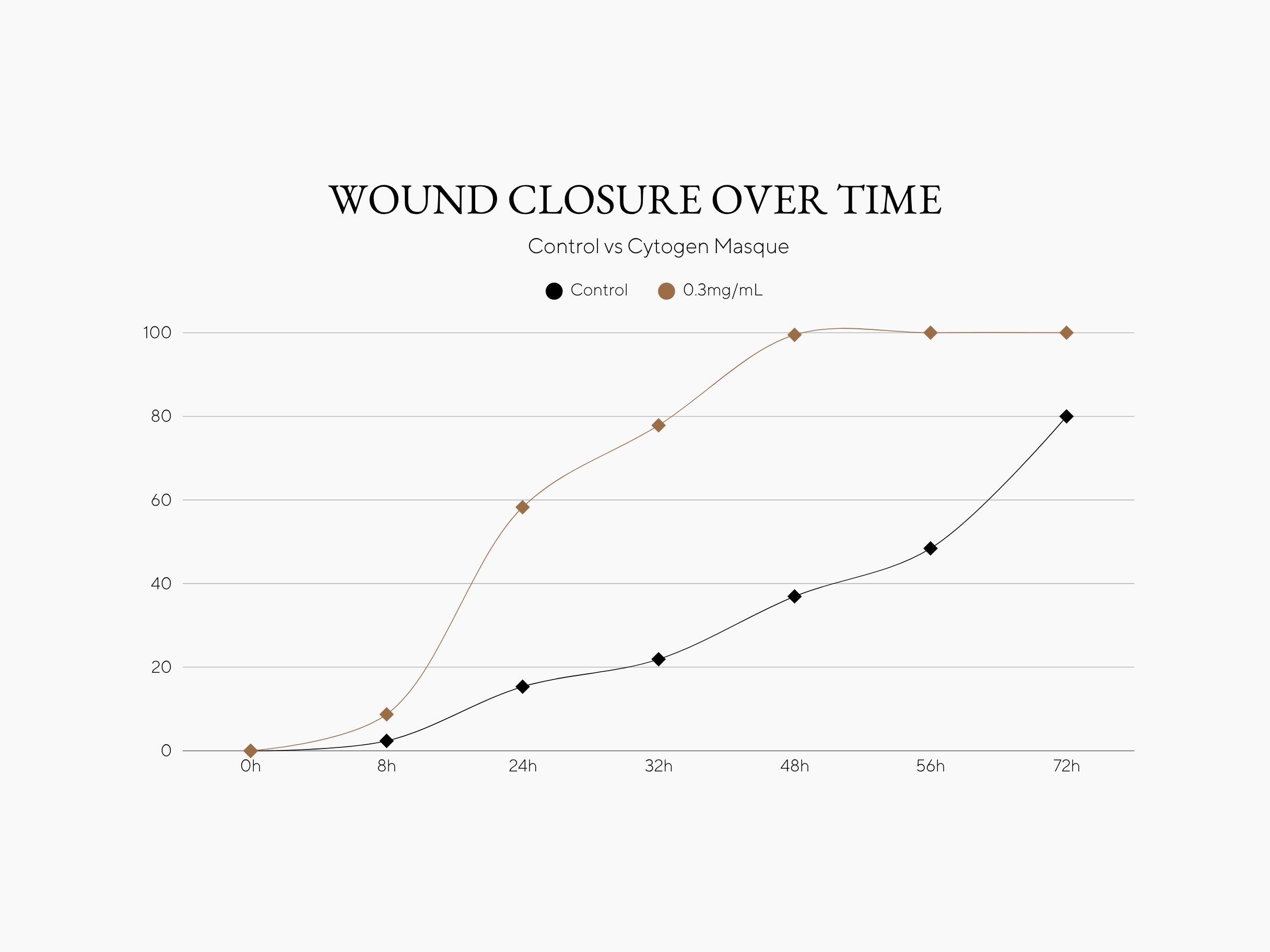
CYTOGEN Masque accelerated keratinocyte gap closure in a standard scratch assay, reaching near-complete closure as early as 32 hours at the lowest tested dose (0.3 mg/mL).
In practical terms, cells treated with the mask extract closed the gap much faster than without treatment.
In-vitro study in HaCaT keratinocytes.
Microscopic Evidence of Accelerated Healing
These microscopy images from the study visually demonstrate how rapidly CYTOGEN Masque promotes keratinocyte migration compared to untreated controls.


Key Findings
- Lowest dose tested (0.3mg/mL) reached near-complete closure by 32 hours.
- Higher doses showed progressively smaller effects - an inverse trend often seen in vitro and worth dose-optimization.
- Controls closed ~79% by 72 hours, confirming baseline migration speed.
Data form HaCaT keratinocyte scratch assay; 7 timepoints over 72h.
EXPONENTIAL WOUND HEALING RECORD
| Concentration | T1 (8h) | T2 (24h) | T3 (32h) | T4 (48h) | T5 (56h) | T6 (72h) |
|---|---|---|---|---|---|---|
| 0.3 mg/ml | +205% | +342% | +314% | +171% | +122% | +27% |
| 0.75 mg/ml | +225% | +256% | +226% | +121% | +97% | +19% |
| 1.5 mg/ml | +138% | +165% | +152% | +81% | +58% | +1% |
How We Tested
Why it matters
- When the monolayer of keratinocytes was “wounded,” Cytogen-treated cells moved significantly faster to close the gap compared to untreated controls.
- At the lowest concentration tested (0.3 mg/ml), Cytogen produced the most potent regenerative activity, outperforming higher concentrations and approaching the performance of HB‑EGF, a known epidermal growth factor used as a positive control.
Microscopic Image Analysis

(NC - t0h) Initial wound area before treatment. A clean, wide gap between keratinocyte layers.

(NC - t24h) Untreated control cells show minimal migration into the wound space after 24 hours.

(PC - t24h) HB-EGF, a known growth factor, shows robust wound closure activity.

(C1 - t24h) Cytogen demonstrates strong wound-healing activity with significant closure approaching the positive control.

(C2 - t24h) Noticeable cell migration into the wound area, outperforming untreated control.

(C3 - t24h) Accelerated healing still present, with visible narrowing of the wound gap.
3D Image Analysis via Quantificare
This section visually compares T-0 (immediately after procedure) with T-15 minutes post-application of the CYTOGEN Mask, highlighting the rapid soothing power of the formula.
The before-and-after imagery reveals a clear reduction in inflammation, redness, and surface irritation, demonstrating how quickly the mask calms compromised skin. The QuantifiCare 3D texture maps and color-coded vascular readings provide an objective, measurable view of the skin’s response—offering undeniable proof of immediate relief and barrier support.


In-Clinic Before and Afters
- Patients experience immediate relief and lasting comfort.
- After just one application, skin appears calmer, more hydrated, and visibly renewed.
- With continued use, barrier strength and resilience visibly improve.
Actual patient treated with a combination of Moxi® fractional laser and a 15-minute CYTOGEN Masque application. Results shown immediately after use. Individual results may vary.
Glossary
Experience CYTOGEN Masque
CYTOGEN Masque moves beyond superficial radiance. It’s a barrier reset rooted in regenerative science — helping skin recover, renew, and build lasting resilience.